
Study Notes
Overview
Welcome to the fascinating world of Cell Biology, the absolute foundation of your AQA GCSE Combined Science course. This topic introduces you to the fundamental units of life, exploring what they're made of and how they work. Understanding cells is crucial because it underpins almost every other topic in biology, from how our bodies function to how ecosystems are structured. In the exam, you can expect a mix of questions: short-answer definitions, labelling diagrams, calculations involving microscopes, and longer 6-mark questions asking you to compare cell types or explain complex processes like mitosis. Mastering this topic early will give you a huge advantage.
Key Concepts
Concept 1: Prokaryotic and Eukaryotic Cells
The first major division you need to understand is between two types of cells: prokaryotic and eukaryotic. It's a fundamental concept that examiners love to test.
-
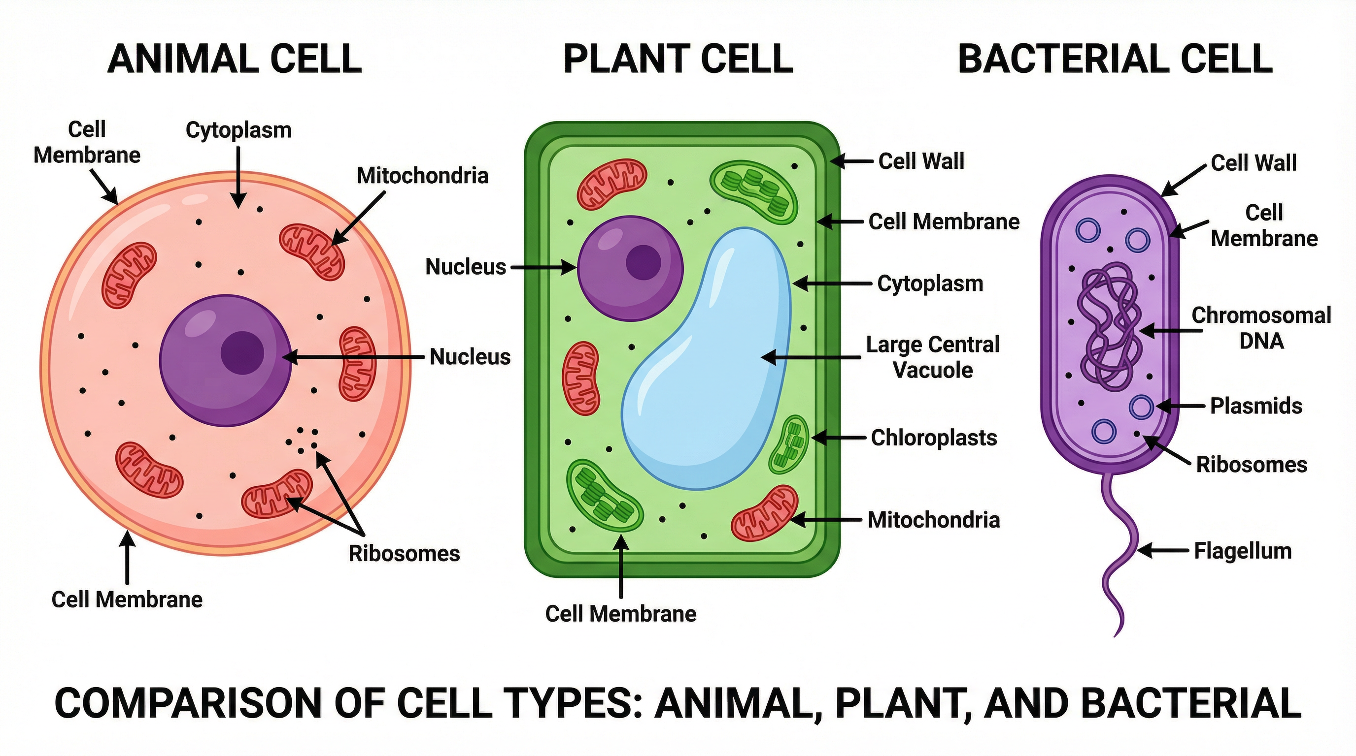
Prokaryotic Cells: These are the simplest and oldest types of cells. Think of bacteria. Their key feature is their simplicity: they have no nucleus. Their genetic material (a single loop of DNA) floats freely in the cytoplasm. Some also have smaller rings of DNA called plasmids. They are much smaller than eukaryotic cells.
-
Eukaryotic Cells: These are more complex and make up all the other forms of life, including plants, animals, fungi, and protists. Their defining feature is that they have a nucleus, which contains the cell's genetic material (DNA) organised into chromosomes. They also contain many other membrane-bound compartments called organelles, each with a specific function.
Concept 2: Animal and Plant Cell Structures
Within the eukaryotes, you need to know the detailed structures of animal and plant cells and be able to compare them.
| Organelle | Animal Cell | Plant Cell | Function | Examiner's Tip |
|---|---|---|---|---|
| Nucleus | Yes | Yes | Contains the genetic material (DNA) and controls the cell's activities. | A key mark-winning phrase is "controls the cell's activities". |
| Cytoplasm | Yes | Yes | A jelly-like substance where most chemical reactions happen. | Don't just say "holds everything"; specify it's the site of chemical reactions. |
| Cell Membrane | Yes | Yes | Controls the movement of substances into and out of the cell. | Use the term "selectively permeable" or "partially permeable" for extra credit. |
| Mitochondria | Yes | Yes | Where aerobic respiration occurs, releasing energy for the cell. | Crucially, they release energy, they do not make or produce it. This is a common mistake. |
| Ribosomes | Yes | Yes | The site of protein synthesis. | Tiny structures, often shown as dots on diagrams. |
| Cell Wall | No | Yes | Made of cellulose, it supports the cell and strengthens it. | Fully permeable. Don't confuse it with the cell membrane! |
| Chloroplasts | No | Yes | Absorb light energy for photosynthesis. Contain a green pigment called chlorophyll. | Found in the green parts of a plant, like leaves and stem. |
| Permanent Vacuole | No | Yes | Contains cell sap (a solution of sugar and salts). Helps to support the cell. | When full, it pushes the cytoplasm against the cell wall, making the cell turgid. |
Concept 3: Cell Specialisation
In multicellular organisms, cells differentiate to become specialised. This means they develop specific features to help them perform a particular job. You need to know several examples for both animals and plants.
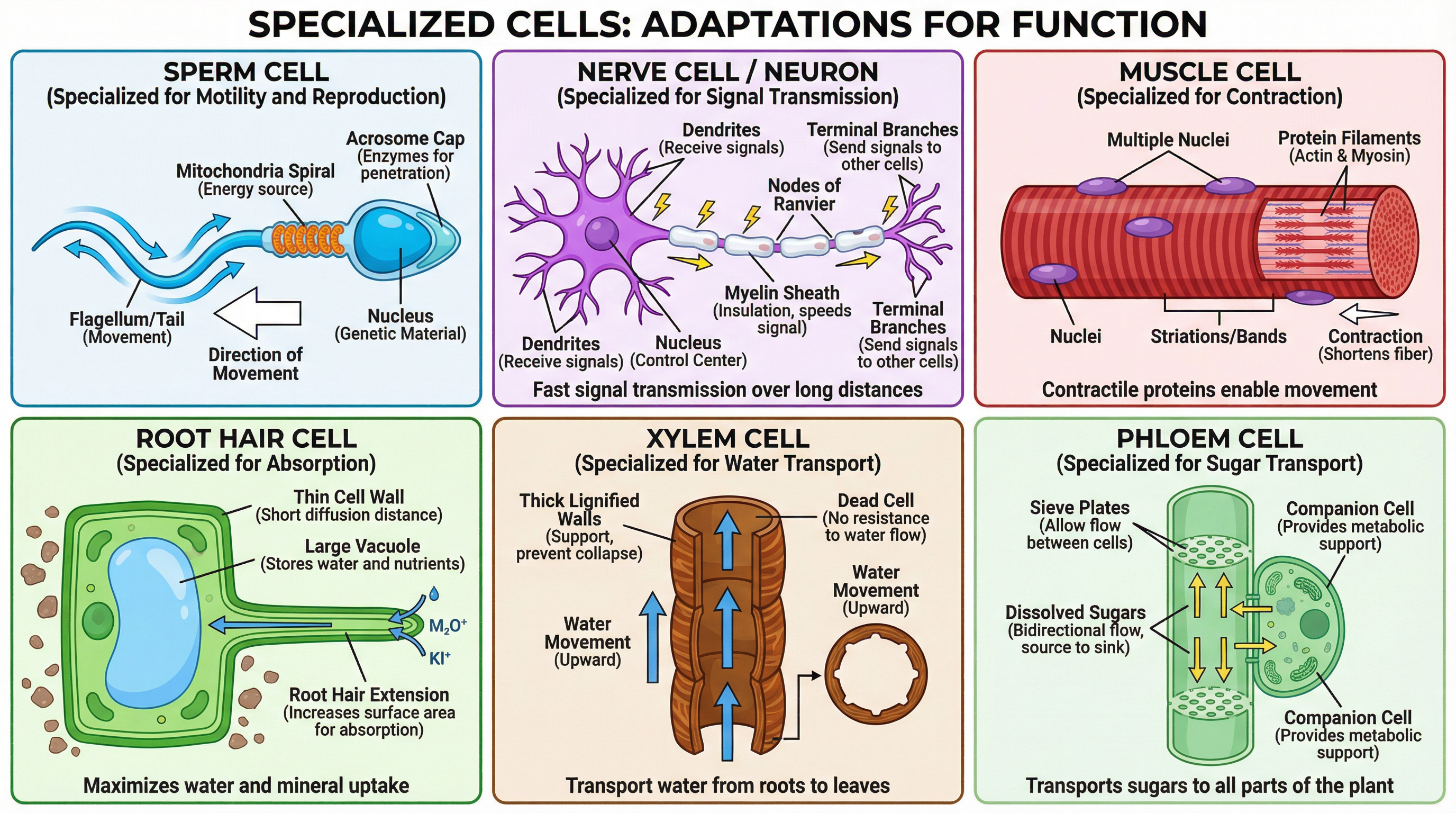
- Sperm Cell: Specialised for reproduction. Adaptations: Long tail (flagellum) for swimming; many mitochondria to release energy; acrosome in the head contains enzymes to digest the egg membrane.
- Nerve Cell (Neuron): Specialised for rapid signalling. Adaptations: Long axon to carry impulses over long distances; branched dendrites to connect with other neurons.
- Muscle Cell: Specialised for contraction. Adaptations: Long cells that can shorten; contain lots of mitochondria for energy.
- Root Hair Cell: Specialised for absorbing water and minerals. Adaptations: Large surface area to increase the rate of absorption.
- Xylem and Phloem Cells: Specialised for transport in plants. Xylem are hollow tubes that transport water. Phloem transport dissolved sugars.
Concept 4: Microscopy and Magnification
Microscopes allow us to see cells. You need to understand the difference between light and electron microscopes and be able to perform magnification calculations.
- Light Microscope: Uses light and lenses. Lower magnification and resolution. Used in schools.
- Electron Microscope: Uses a beam of electrons. Much higher magnification and resolution, allowing us to see sub-cellular structures in detail.
Magnification CalculationThis is a guaranteed mark-winner if you learn the formula and are careful with units.
Magnification = Image Size / Actual Size
Crucial Unit Conversions:
- 1 millimetre (mm) = 1000 micrometres (µm)
- 1 micrometre (µm) = 1000 nanometres (nm)
Examiner's Trap: They will give you the image size in mm and the actual size in µm. You MUST convert them to the same unit before calculating. The easiest way is to convert the mm value to µm by multiplying by 1000.
Concept 5: Mitosis and the Cell Cycle
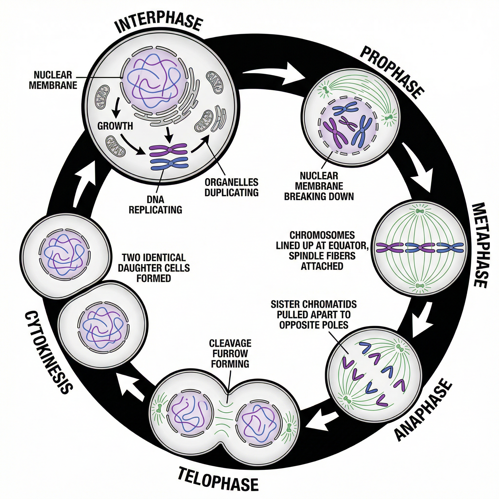
Multicellular organisms grow and repair tissues by making more cells. This is achieved through the cell cycle, which includes a process called mitosis.
The Cell Cycle: A two-stage process.
- Growth and DNA Replication: The cell grows larger. The number of sub-cellular structures (like mitochondria and ribosomes) increases. Crucially, the DNA replicates to form two copies of each chromosome.
- Mitosis: The nucleus divides. One set of chromosomes is pulled to each end of the cell.
Following mitosis, the cytoplasm and cell membrane divide to form two genetically identical daughter cells.
Mitosis is essential for:
- Growth of the organism.
- Repairing damaged tissues.
- Asexual reproduction in some organisms.
Mathematical/Scientific Relationships
Magnification Formula
- Formula:
Magnification = Image Size / Actual Size - How to remember: Use the I-A-M formula triangle. Cover the value you want to find.
- Status: Must memorise.
Unit Conversions
- 1 mm = 1000 µm
- 1 µm = 1000 nm
- Status: Must memorise and be able to apply confidently.
Required Practical: Using a Light Microscope
Apparatus:
- Light microscope
- Glass slide and coverslip
- Pipette
- Onion
- Forceps
- Iodine solution (stain)
Method:
- Add a drop of water to the middle of a clean slide.
- Use forceps to peel off a thin layer of epidermal tissue from the inside of an onion.
- Place the onion tissue into the water on the slide.
- Add a drop of iodine solution. This is a stain used to make the sub-cellular structures (like the nucleus) visible.
- Carefully place a coverslip on top. Try to avoid air bubbles.
- Clip the slide onto the microscope stage.
- Select the lowest-power objective lens.
- Use the coarse adjustment knob to move the stage up until the image is roughly in focus.
- Use the fine adjustment knob to bring the image into a clear focus.
- To see the cells in greater detail, switch to a higher-power objective lens and refocus using the fine adjustment knob.
Expected Results: You will see a grid-like pattern of rectangular cells. The cell walls, cytoplasm, and a dark-stained nucleus should be visible.
How Examiners Test It: They may ask you to describe the method, explain the purpose of the stain, or calculate the magnification of a drawing you make.