Study Notes
Overview
The study of neurons and synaptic transmission is a cornerstone of the AQA A-Level Psychology (Biopsychology) specification. It explores the structure and function of the nervous system at a microscopic level, detailing how information is passed through the body via a combination of electrical and chemical signals. For candidates, this is not a topic for vague descriptions; examiners award marks for precise, sequential, and technically accurate explanations. A firm grasp of the action potential, the roles of different neurons, the sequence of synaptic transmission, and the crucial process of summation is essential. This guide will break down these complex processes into manageable sections, providing the level of detail required to achieve top marks and highlighting the common pitfalls that cost candidates valuable credit.
The Building Blocks: Structure and Types of Neuron
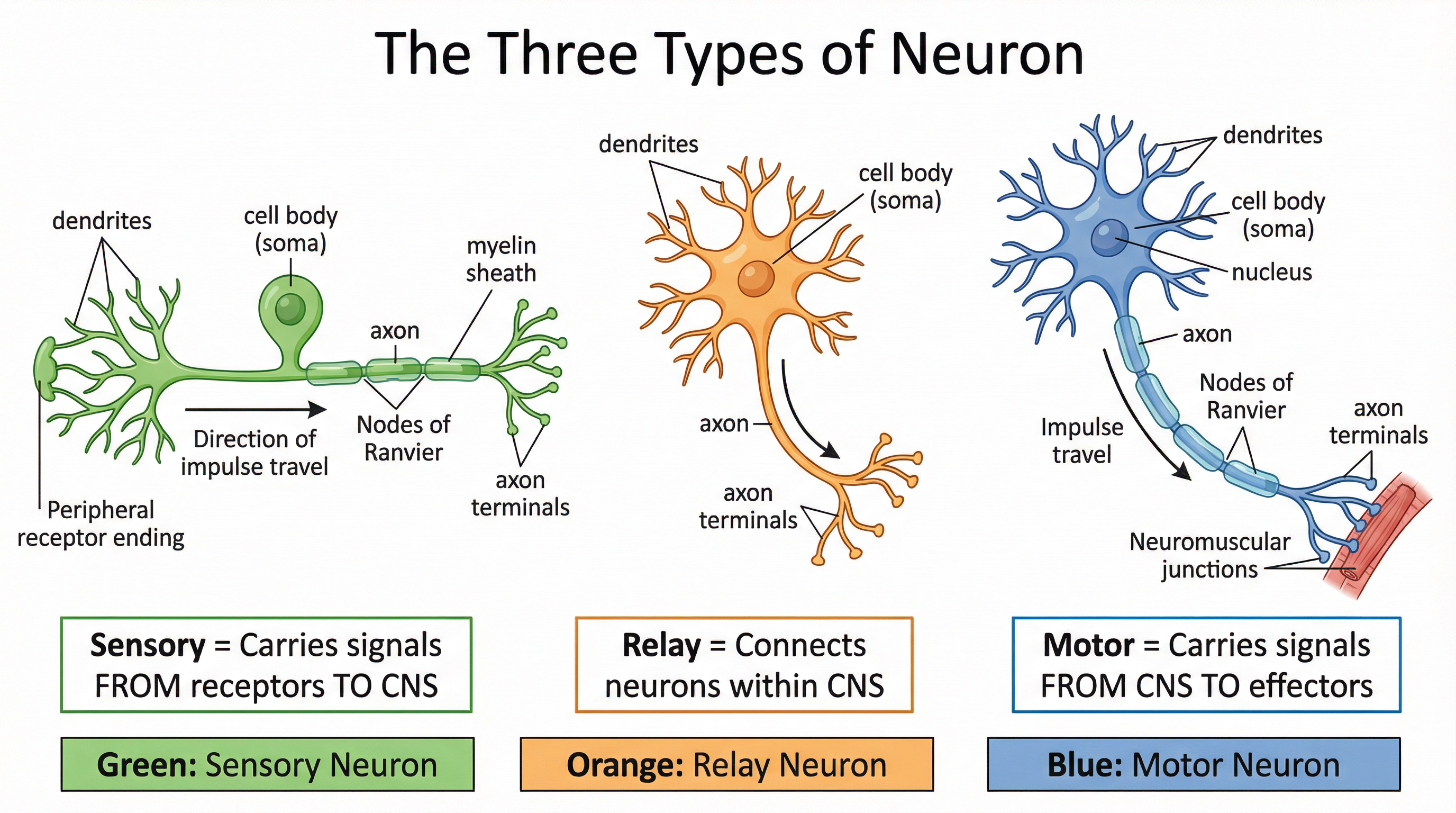
Neurons are the primary cells of the nervous system, responsible for transmitting nerve impulses. To gain credit, candidates must be able to identify and differentiate between the three main types.
| Neuron Type | Function | Key Structural Features |
|---|---|---|
| Sensory Neuron | Transmits nerve impulses from sensory receptors (e.g., in the skin, eyes) towards the Central Nervous System (CNS). | Long dendrites, short axon. Cell body is often located to one side. |
| Relay Neuron | Transmits nerve impulses between other neurons, typically as part of a pathway within the CNS. | Short dendrites, short axon. Entirely contained within the CNS. |
| Motor Neuron | Transmits nerve impulses from the CNS to an effector, such as a muscle or gland. | Short dendrites, long axon. Cell body is at one end. |
The Electrical Impulse: The Action Potential
Communication within a neuron is an electrical process. The action potential is a brief, powerful electrical charge that travels down the axon. Candidates must understand the sequence of ionic exchange that creates this impulse.
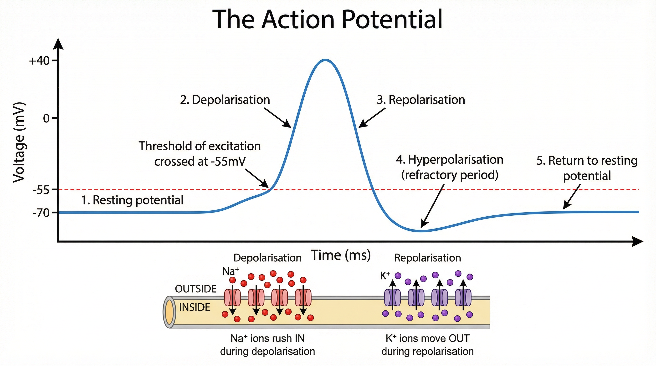
- Resting State: The neuron is not firing. The inside of the axon is negatively charged relative to the outside (around -70mV). This is the resting potential.
- Depolarisation: When stimulated, sodium ion (Na+) channels open and Na+ ions flood into the axon, causing the inside to become positively charged (around +40mV). This requires the initial stimulation to reach the threshold of excitation (approx. -55mV).
- Repolarisation: Following the spike in charge, sodium channels close and potassium ion (K+) channels open. K+ ions flood out of the axon, returning the inside to a negative charge.
- Hyperpolarisation: The axon briefly becomes more negative than its resting state. This is the refractory period, during which it cannot fire again, ensuring the impulse travels in one direction.
- Return to Resting State: The sodium-potassium pump actively restores the balance of ions, returning the neuron to its -70mV resting potential.
This process follows the all-or-nothing law: the size of the action potential is always the same. It either fires completely or not at all.
The Chemical Bridge: Synaptic Transmission
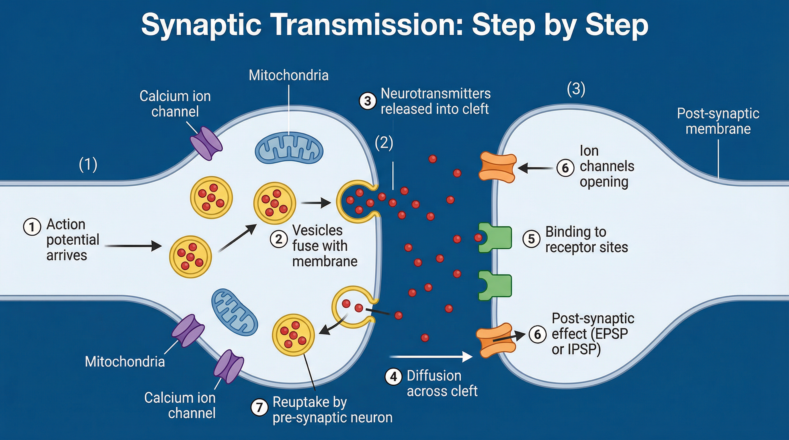
Neurons do not physically touch. The gap between the pre-synaptic neuron and the post-synaptic neuron is the synaptic cleft. Communication across this gap is a chemical process. Marks are awarded for a clear, sequential description.
The Sequence of Synaptic Transmission:
- Arrival of Action Potential: The electrical impulse reaches the pre-synaptic terminal.
- Vesicle Release: The action potential triggers voltage-gated calcium channels to open. Calcium ions (Ca2+) flood into the terminal, causing synaptic vesicles (sacs containing neurotransmitters) to fuse with the pre-synaptic membrane.
- Diffusion: The neurotransmitters are released into the synaptic cleft and diffuse across the gap.
- Receptor Binding: Neurotransmitter molecules bind to complementary receptor sites on the post-synaptic membrane. This is often described using the 'lock and key' analogy.
- Post-synaptic Effect: This binding causes ion channels to open on the post-synaptic neuron, resulting in either an Excitatory Post-synaptic Potential (EPSP), which makes the neuron more likely to fire, or an Inhibitory Post-synaptic Potential (IPSP), which makes it less likely to fire.
- Termination: The neurotransmitter is removed from the synapse to prevent continuous stimulation. This occurs via re-uptake (the pre-synaptic neuron reabsorbs the neurotransmitter) or enzymatic degradation (an enzyme breaks down the neurotransmitter in the cleft).
The Decision to Fire: Summation
A post-synaptic neuron receives thousands of inputs. Summation is the process of adding up all the excitatory and inhibitory signals. If the net effect of the signals reaches the threshold of excitation (-55mV), the post-synaptic neuron will fire an action potential.
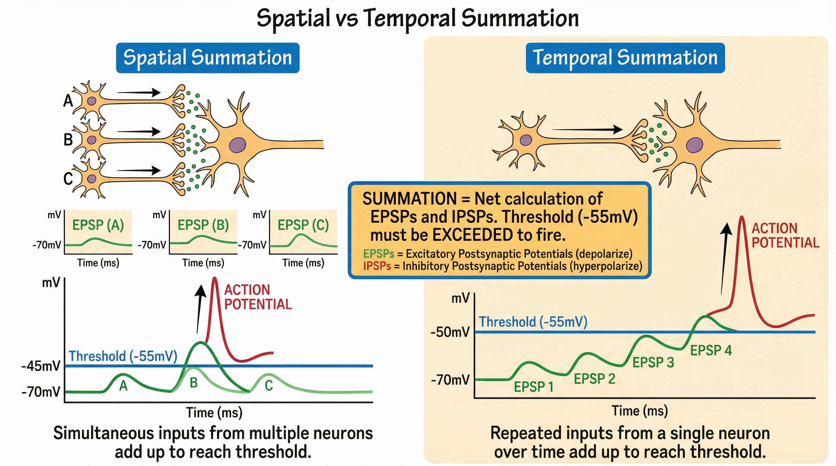
- Temporal Summation: A single pre-synaptic neuron fires repeatedly in quick succession, building up the post-synaptic potential over time.
- Spatial Summation: Multiple pre-synaptic neurons fire simultaneously onto the same post-synaptic neuron, combining their effects.