Study Notes
Overview
Ultrasound sits within the Waves section of the OCR GCSE Physics specification (Topic 4.9) and is one of the most reliably examined topics at both Foundation and Higher tier. It bridges fundamental wave physics — longitudinal waves, reflection, and the wave equation — with real-world applications in medicine and oceanography that examiners frequently use as novel contexts for AO2 application questions.
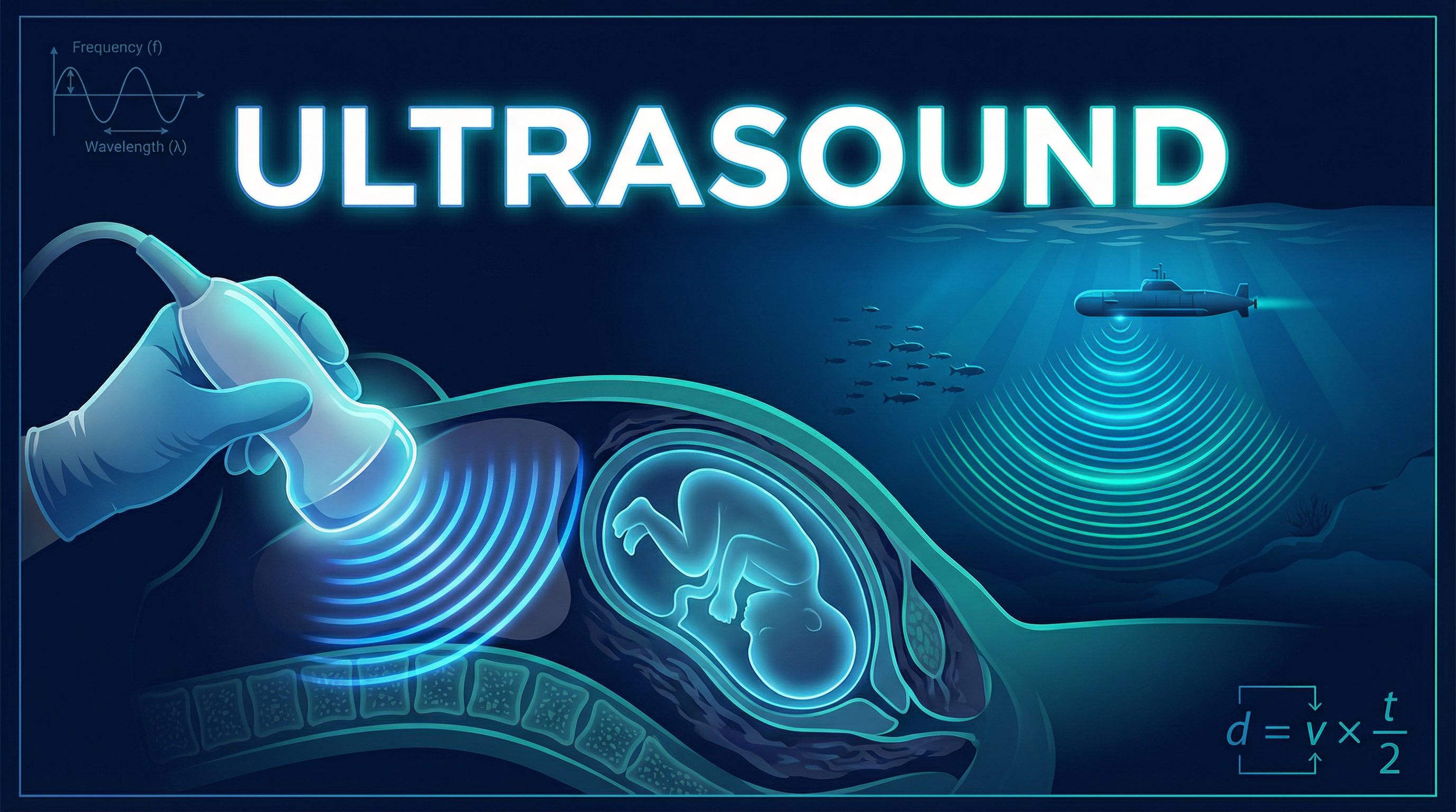
At its core, this topic asks candidates to understand that ultrasound is simply sound with a frequency too high for humans to hear (above 20,000 Hz), and that its usefulness in imaging arises from a single elegant physical principle: when a wave crosses a boundary between two materials of different densities, part of it reflects back. By measuring how long these reflections take to return, machines can calculate distances and construct images of internal structures.
Exam questions on this topic typically fall into three categories: short-answer definition questions (1–2 marks, AO1), calculation questions involving echo sounding (3–4 marks, AO2), and extended comparison or evaluation questions comparing ultrasound with X-rays (4–6 marks, AO1/AO2/AO3). Candidates who understand the underlying physics — rather than memorising isolated facts — consistently outperform those who do not.
Key Concepts
Concept 1: What is Ultrasound?
Ultrasound is defined as sound waves with a frequency greater than 20,000 Hz (20 kHz). This is the upper limit of the human audible range. The human ear can detect frequencies between approximately 20 Hz (lower limit, below which is infrasound) and 20,000 Hz. Ultrasound lies above this range and is therefore inaudible to humans, though many animals — including dogs, bats, and dolphins — can both produce and detect it.
Critically, ultrasound is a longitudinal wave. In a longitudinal wave, the particles of the medium vibrate parallel to the direction of wave propagation, creating alternating regions of compression (where particles are pushed together) and rarefaction (where particles are spread apart). This is in contrast to transverse waves such as light or water waves, where particle vibration is perpendicular to the direction of travel. OCR mark schemes regularly award a mark specifically for identifying ultrasound as longitudinal, so candidates must not confuse the two types.
Like all mechanical waves, ultrasound requires a medium through which to travel — it cannot propagate through a vacuum. Its speed depends on the medium: approximately 340 m/s in air, around 1,500 m/s in water and soft tissue, and higher still in denser solids like bone.
Analogy: Think of ultrasound like an extremely rapid series of claps. Each clap sends a pressure pulse through the air (or tissue). The frequency is simply how many of these pulses occur per second — for ultrasound, more than 20,000 per second, far too fast for our ears to register as separate events.
Concept 2: Partial Reflection at Boundaries
The entire principle of ultrasound imaging rests on what happens when a wave encounters a boundary between two media of different densities.
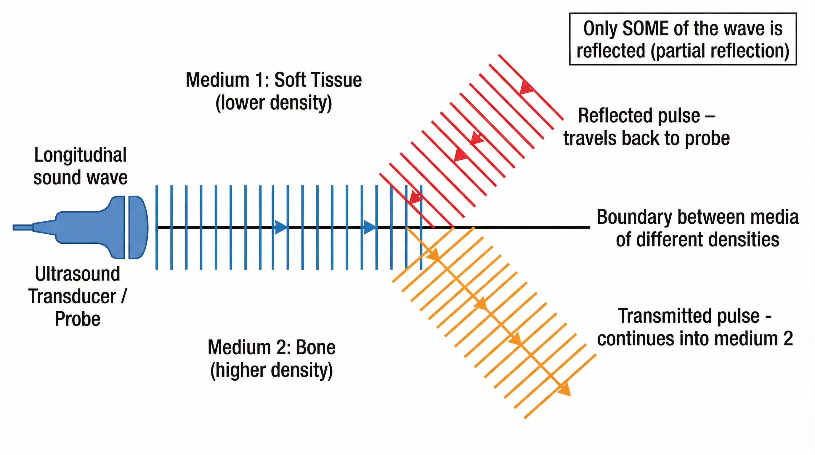
When an ultrasound pulse travelling through one medium (for example, soft tissue with a certain density) reaches the interface with a second medium of a different density (for example, bone, which is considerably denser), the wave does not simply pass straight through. Instead, it splits:
- A portion of the wave is reflected back towards the source (the probe). This is the reflected pulse, or echo.
- The remainder of the wave is transmitted into the second medium, continuing forward (though it may change speed and direction slightly — this is refraction).
This phenomenon is called partial reflection. The word partial is absolutely critical and must appear in any exam answer describing this process. Saying the wave simply 'bounces off' will not earn marks; the mark scheme requires candidates to state that some of the wave is reflected and some is transmitted.
The amount of reflection depends on the difference in density between the two media. The greater the contrast in density, the stronger the reflected pulse. This is why ultrasound produces clear images at boundaries between soft tissue and bone (large density difference) but may struggle to distinguish between two tissues of very similar density.
In a medical scan, the probe acts as both the transmitter (emitting the pulse) and the receiver (detecting the returning echo). The machine processes each reflected pulse and its time delay to determine the depth of each boundary, building up a two-dimensional image from many such measurements.
Concept 3: Echo Sounding and the Distance Calculation
The mathematical heart of this topic is the echo-sounding calculation. The fundamental relationship is the wave equation:
distance = speed × time, or d = v × tHowever, in all echo-sounding contexts — whether sonar at sea or medical ultrasound — there is a crucial modification: the time measured is the total time for the pulse to travel to the boundary AND back. The pulse makes a return journey. Therefore, the time recorded represents twice the actual distance to the boundary.
The correct formula for depth in echo sounding is:
depth = (speed × total time) ÷ 2, or equivalently, d = v × (t/2)
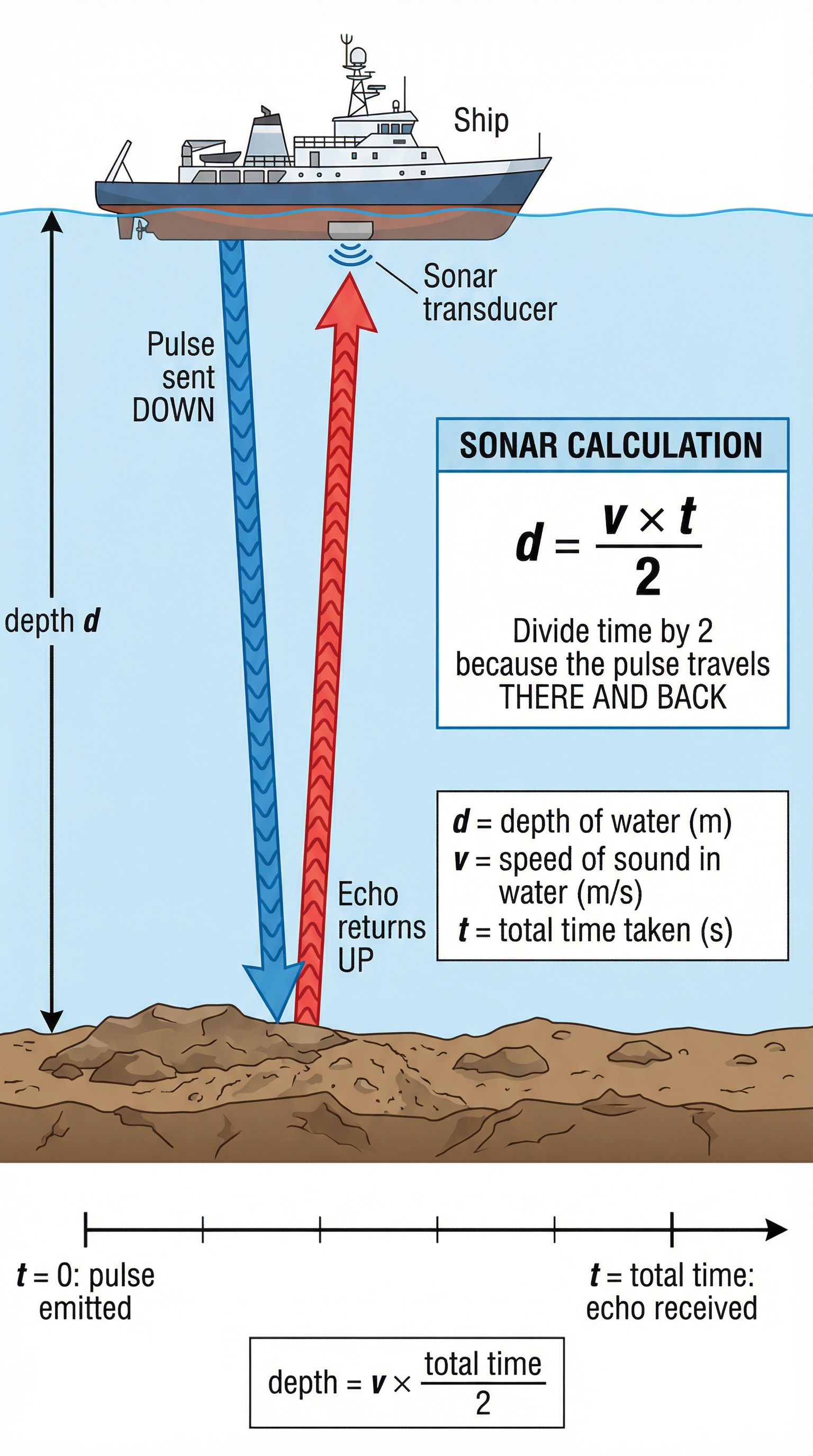
This halving of the time is the single most tested — and most frequently missed — element of this topic. OCR examiners report that failing to divide by two is the most common error in ultrasound calculation questions, resulting in a depth value exactly double the correct answer.
Worked Calculation:
A ship uses sonar to map the ocean floor. A pulse is emitted and the echo returns after 0.4 s. The speed of sound in seawater is 1,500 m/s. Find the depth.
- Step 1: Identify the formula: d = v × t/2
- Step 2: Substitute: d = 1,500 × (0.4 ÷ 2) = 1,500 × 0.2
- Step 3: Calculate: d = 300 m
The depth of the ocean floor is 300 m.
Concept 4: Applications of Ultrasound
Medical Imaging: Ultrasound scanning is used extensively in medicine to image soft tissue structures — organs, blood vessels, tendons, and developing foetuses. Because ultrasound is non-ionising, it does not carry the risk of DNA damage associated with ionising radiation such as X-rays or gamma rays. This makes it the preferred imaging modality during pregnancy, where repeated exposure to ionising radiation could harm the developing foetus.
The probe is placed on the skin (with a gel to ensure good acoustic contact and prevent air gaps that would cause total reflection) and emits pulses that penetrate the body. Reflected pulses from each tissue boundary return to the probe at different times, and the machine converts these time delays into a real-time image on screen.
Sonar (Sound Navigation and Ranging): Ships and submarines use sonar to measure water depth, detect underwater obstacles, and locate fish shoals. The same echo-sounding principle applies: a pulse is emitted downward, and the time for the echo to return from the seabed is used to calculate depth.
Industrial Applications (Higher tier context): Ultrasound is also used in non-destructive testing of materials — for example, detecting cracks inside metal components. A crack creates a boundary (metal-to-air interface) that reflects the ultrasound pulse, revealing the defect without cutting the material open.
Concept 5: Comparing Ultrasound and X-rays
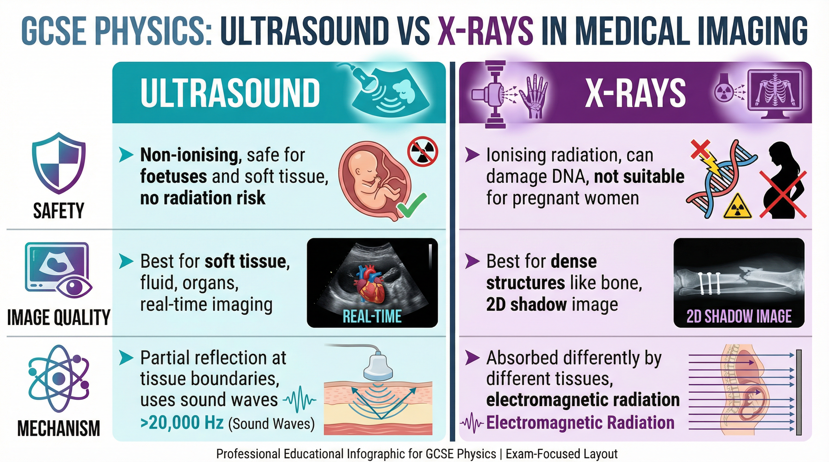
A common 6-mark question asks candidates to compare ultrasound and X-ray imaging. The OCR mark scheme for such questions typically awards marks under three headings: Safety, Image Quality, and Mechanism of Action. Candidates should structure their answer accordingly.
| Feature | Ultrasound | X-rays |
|---|---|---|
| Wave type | Longitudinal sound wave | Transverse electromagnetic wave |
| Frequency | > 20,000 Hz | ~10^18 Hz |
| Ionising? | No — non-ionising | Yes — ionising |
| Safe in pregnancy? | Yes | No |
| Best for | Soft tissue, organs, foetuses | Bone, dense structures |
| Mechanism | Partial reflection at boundaries | Differential absorption by tissues |
| Image type | Real-time, 2D cross-section | 2D shadow image |
Mathematical Relationships
Core Formula: Wave Equation
d = v × t (distance = speed × time)
- d = distance (metres, m) — Must memorise; not on OCR formula sheet
- v = wave speed (metres per second, m/s)
- t = time (seconds, s)
Echo Sounding Modification
d = v × t/2 (depth = speed × half the total time)
- Used whenever the time given represents a return journey (pulse travels to boundary and back)
- Always check: is the time one-way or two-way? In exam questions, it is almost always two-way.
Frequency and the Audible Range
- Human hearing range: 20 Hz to 20,000 Hz
- Infrasound: < 20 Hz
- Ultrasound: > 20,000 Hz (20 kHz)
Typical Speeds of Sound (for context — values given in exam)
| Medium | Approximate Speed |
|---|---|
| Air (20°C) | ~340 m/s |
| Water / soft tissue | ~1,500 m/s |
| Bone | ~3,000–4,000 m/s |
Practical Applications and Required Practicals
Ultrasound does not have a dedicated required practical in the OCR GCSE specification, but the topic is frequently assessed through data-analysis questions using oscilloscope traces or time-distance graphs showing reflected pulses. Candidates should be able to:
- Read a time axis on an oscilloscope trace to identify the time delay between the emitted pulse and the reflected echo.
- Identify multiple reflections — a trace may show two reflected pulses (from two different boundaries at different depths), and candidates must correctly assign each echo to its corresponding boundary.
- Calculate depth from the time delay and given wave speed.
- Interpret what a longer time delay means — a greater time delay indicates a deeper boundary.
Graph skills: If plotting depth against time delay, the relationship should be directly proportional (a straight line through the origin), since d = v × t/2 and v is constant.